| Strain Name |
C57BL/6-Egfrtm2(EGFR)Bcgen/Bcgen
|
Common Name | B-hEGFR mice |
| Background | C57BL/6 | Catalog number | 120771 |
| Aliases |
EGFR, ERBB, ERBB1, ERRP, HER1, NISBD2, PIG61, mENA, epidermal growth factor receptor |
||
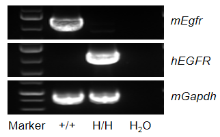
Strain specific analysis of EGFR gene expression in wild type (WT) mice and B-hEGFR mice by RT-PCR. Mouse Egfr mRNA was detectable only in liver of WT mice (+/+). Human EGFR mRNA was detectable only in homozygous B-hEGFR mice (H/H) but not in WT mice (+/+).

Immunohistochemical (IHC) analysis of EGFR expression in homozygous B-hEGFR mice. The liver and kidney were collected from WT and homozygous B-hEGFR mice (H/H) and analyzed by IHC with anti-EGFR antibody. Mouse EGFR was detectable in WT mice, and human EGFR was detectable in homozygous B-hEGFR mice. The arrow indicates tissue cells with positive EGFR staining (brown).
Protein expression profile of EGFR
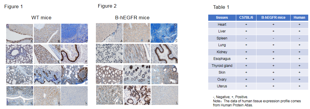
Immunohistochemical (IHC) analysis of EGFR protein expression in wild-type (WT) mice and B-hEGFR mice. Ten major tissues were collected from WT mice and homozygous B-hEGFR mice (2 females, 8 weeks-old), and analyzed by IHC with anti-EGFR antibodies. Humanized EGFR was detected in the heart (A), liver (B), lung (D), kidney (E), esophagus (F), thyroid (G), skin (H), ovary (I) and uterus (J) of homozygous B-hEGFR mice (Figure 2) and human EGFR positive control (K), but not in spleen (C) and human EGFR negative control (L). This is similar to the mouse EGFR expression profile of WT mice (Figure 1, Table 1). The results showed that humanized EGFR did not change the expression site of EGFR protein in B-hEGFR mice, and the expression profile of EGFR in B-hEGFR mice was similar to that in normal human tissues (Table 1).
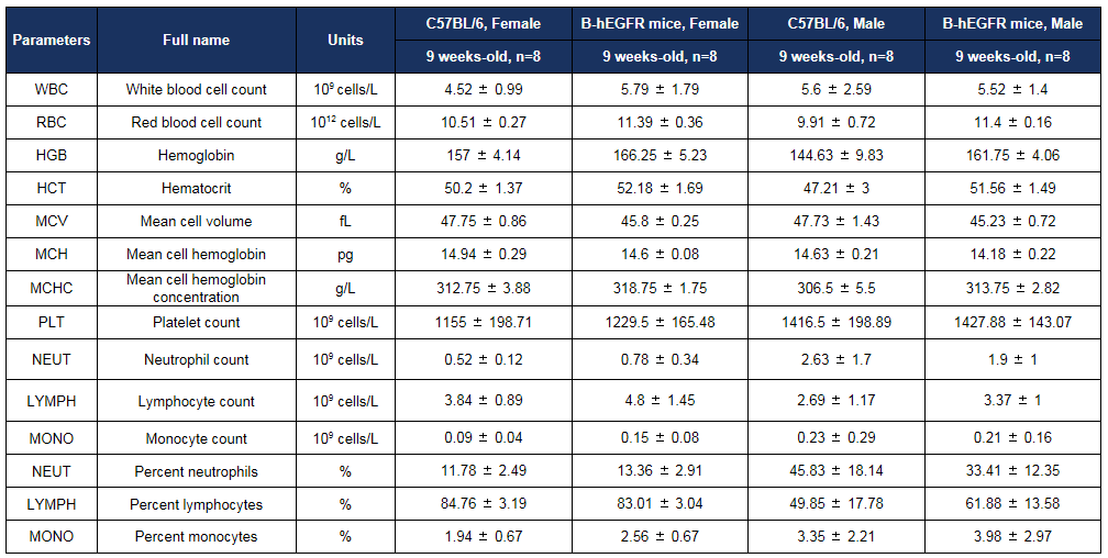
Complete blood count (CBC) of B-hEGFR mice. Values are expressed as mean ± SD.
Biochemistry analysis
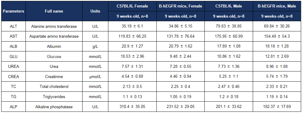
Biochemical test of B-hEGFR mice. Values are expressed as mean ± SD.

Antitumor activity of anti-human EGFR antibody in B-hEGFR mice. (A) Anti-human EGFR antibody inhibited B-Tg(hEGFR) MC38 tumor growth in homozygous B-hEGFR mice. Murine colon cancer B-Tg(hEGFR) MC38 cells were subcutaneously implanted into homozygous B-hEGFR mice (female, 6-8 weeks-old, n=6). Mice were grouped when tumor volume reached approximately 50-150 mm3, at which time they were intravenous injected with anti-human EGFR ADC cetuximab analog-MMAE (in house) indicated in panel. (B) Body weight changes during treatment. As shown in panel A, 30mg/kg anti-human EGFR ADC cetuximab analog-MMAE (in house) treatment group was efficacious in controlling tumor growth in B-hEGFR mice, demonstrating that the B-Tg(hEGFR) MC38 provide a powerful preclinical model for in vivo evaluation of anti-human EGFR antibodies. Values are expressed as mean ± SEM.
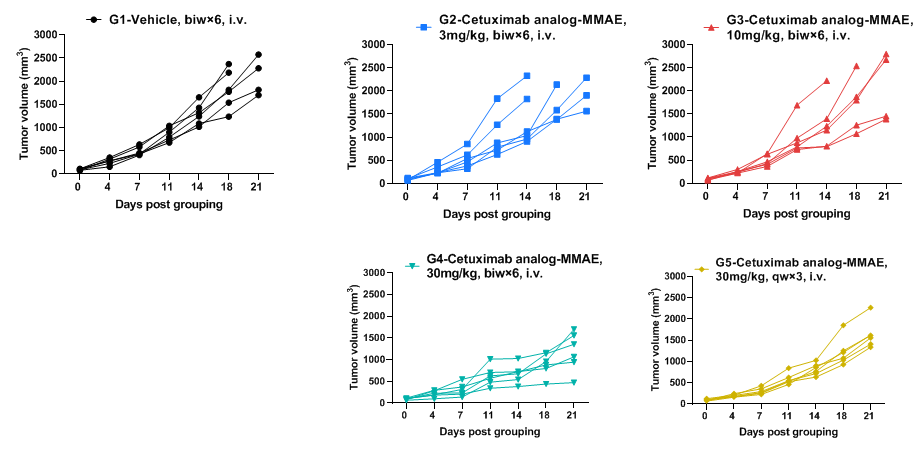
Antitumor activity of anti-human EGFR antibody in B-hEGFR mice. Anti-human EGFR antibody inhibited B-Tg(hEGFR) MC38 tumor growth in homozygous B-hEGFR mice. Murine colon cancer B-Tg(hEGFR) MC38 cells were subcutaneously implanted into homozygous B-hEGFR mice (female, 6-8 weeks-old, n=6). Mice were grouped when tumor volume reached approximately 50-150 mm3, at which time they were intravenous injected with anti-human EGFR ADC cetuximab analog-MMAE (in house) indicated in panel.


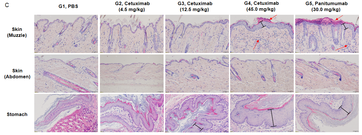
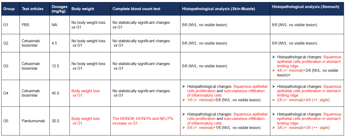
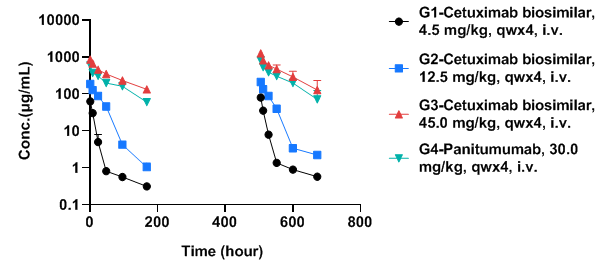
Toxicokinetic analysis of anti-human EGFR antibody in B-hEGFR mice
 苏公网安备:32068402320845号
网站建设:北京分形科技


 010-56967680
 info@bbctg.com.cn